A state of as shown by eeg – Delving into the realm of brain activity, we explore a state of wakefulness as shown by EEG. Electroencephalography (EEG), a non-invasive technique, provides a window into the electrical rhythms of the brain, offering insights into our conscious state.
As we navigate through this topic, we will uncover the distinctive EEG patterns associated with wakefulness, delve into the physiological and cognitive processes that orchestrate our alert state, and explore the diagnostic and monitoring applications of EEG in unraveling neurological conditions.
State of Wakefulness

The state of wakefulness is characterized by high levels of alertness and responsiveness to the environment. It is the state in which we are most actively engaged with the world around us.
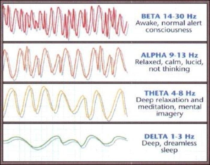
The EEG patterns associated with wakefulness are characterized by low-amplitude, high-frequency waves. These waves are known as beta waves and have a frequency of 13-30 Hz. Beta waves are associated with cognitive processes such as attention, problem-solving, and decision-making.
A state of find the measure of yvz as shown by EEG can provide valuable insights into brain activity. This technique has been used to diagnose various neurological conditions, such as epilepsy and sleep disorders. By measuring the electrical activity of the brain, EEG can help identify abnormalities that may be causing symptoms.
Furthermore, EEG can be used to monitor the effectiveness of certain treatments, such as medication or surgery.
Physiological and Cognitive Processes During Wakefulness
During wakefulness, the brain is highly active. The reticular activating system (RAS) is a network of neurons in the brainstem that is responsible for maintaining wakefulness. The RAS receives input from the sensory organs and sends signals to the cerebral cortex, which is the part of the brain responsible for higher-level cognitive functions.
The RAS also plays a role in regulating sleep-wake cycles. When the RAS is active, we are awake. When the RAS is inactive, we fall asleep.
Sleep Stages
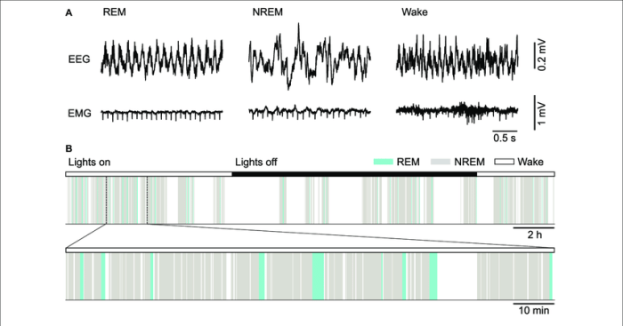
Sleep is a complex process that involves distinct stages, each characterized by specific EEG patterns and physiological changes. As we progress from wakefulness to sleep, our EEG patterns undergo a series of transformations that reflect the varying levels of brain activity.
Non-REM Sleep
- Stage N1:Transition from wakefulness to sleep. EEG shows a mix of alpha and theta waves, with occasional K-complexes and sleep spindles.
- Stage N2:Light sleep. EEG shows more theta waves, along with sleep spindles and K-complexes. Body temperature and heart rate decrease.
- Stage N3:Deep sleep. EEG shows slow delta waves. Breathing and heart rate are at their lowest. Muscle tone is reduced, and it is difficult to wake the individual.
REM Sleep
REM sleep is characterized by rapid eye movements and EEG patterns that resemble those of wakefulness. It is associated with dreaming and memory consolidation.
- EEG patterns:Similar to wakefulness, with low-amplitude, high-frequency beta waves.
- Physiological changes:Muscles are paralyzed, but the eyes move rapidly. Breathing and heart rate increase.
EEG Abnormalities: A State Of As Shown By Eeg

Electroencephalography (EEG) abnormalities are deviations from the normal EEG patterns and can indicate various neurological conditions. Identifying and interpreting these abnormalities is crucial for diagnosis and monitoring of these conditions.
Epilepsy
- Interictal epileptiform discharges (IEDs): Sharp or spike-wave discharges that occur outside of seizures and indicate an increased risk of seizures.
- Ictal EEG patterns: Characteristic patterns that occur during seizures, such as rhythmic discharges or spike-and-wave complexes.
Brain Tumors
- Focal slowing: Slowing of brain activity in a specific area, indicating a tumor or other mass lesion.
- Asymmetrical EEG: Differences in EEG activity between the two hemispheres, suggesting a tumor or other structural abnormality.
Encephalitis
- Diffuse slowing: Generalized slowing of brain activity, indicating inflammation of the brain.
- Periodic lateralized epileptiform discharges (PLEDS): Rhythmic, high-amplitude discharges that occur on one side of the brain, associated with herpes simplex encephalitis.
Creutzfeldt-Jakob Disease (CJD)
- Periodic sharp wave complexes (PSWCs): Characteristic bursts of sharp waves that occur every 1-2 seconds, indicating CJD.
- Triphasic waves: Distinctive waves with three phases, also associated with CJD.
Case Study: Epilepsy Diagnosis
A patient with suspected epilepsy underwent an EEG. The EEG showed interictal epileptiform discharges in the left temporal lobe. This abnormality suggested an increased risk of seizures and helped in diagnosing the patient with epilepsy.
EEG Monitoring

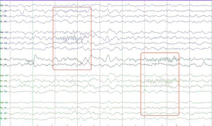
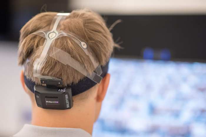
Electroencephalography (EEG) monitoring is a technique that records electrical activity in the brain. It involves placing electrodes on the scalp to detect and amplify the brain’s electrical signals, which are then displayed as a series of waveforms on a computer screen or chart recorder.
EEG monitoring is used to assess brain function and diagnose a variety of neurological conditions, including epilepsy, sleep disorders, and encephalopathy. It can also be used to monitor the effects of anesthesia and sedation, and to evaluate brain death.
Principles and Techniques of EEG Monitoring
EEG monitoring is based on the principle that the brain produces electrical signals that can be detected and recorded. These signals are generated by the synchronized activity of large populations of neurons in the brain. The frequency and amplitude of the EEG signals vary depending on the state of the brain, such as whether the person is awake, asleep, or in a state of altered consciousness.
EEG monitoring is typically performed using a set of electrodes that are placed on the scalp. The electrodes are connected to an amplifier, which amplifies the electrical signals and sends them to a computer or chart recorder. The computer or chart recorder then displays the EEG signals as a series of waveforms.
Applications of EEG Monitoring in Clinical Settings
EEG monitoring is used in a variety of clinical settings, including:
- Epilepsy:EEG monitoring is used to diagnose epilepsy and to classify the type of seizures that a person is experiencing. It can also be used to monitor the effectiveness of anti-epileptic medications.
- Sleep disorders:EEG monitoring is used to diagnose sleep disorders, such as insomnia, narcolepsy, and sleep apnea. It can also be used to monitor the effectiveness of sleep medications.
- Encephalopathy:EEG monitoring is used to diagnose encephalopathy, which is a condition that affects brain function. Encephalopathy can be caused by a variety of factors, including head injuries, strokes, and infections.
- Anesthesia and sedation:EEG monitoring is used to monitor the effects of anesthesia and sedation. It can help to ensure that the patient is adequately anesthetized or sedated, and that there are no adverse effects on brain function.
- Brain death:EEG monitoring is used to evaluate brain death. A person is considered brain dead if there is no electrical activity in the brain for a period of time.
Advantages and Limitations of EEG Monitoring
EEG monitoring has a number of advantages over other diagnostic methods, including:
- It is a non-invasive procedure that does not require the use of radiation.
- It can be performed in a variety of settings, including hospitals, clinics, and even at home.
- It provides a continuous record of brain activity, which can be helpful in diagnosing and monitoring neurological conditions.
However, EEG monitoring also has some limitations, including:
- It can be difficult to interpret EEG signals, especially in patients with complex neurological conditions.
- EEG monitoring can be time-consuming and expensive.
- EEG monitoring is not always able to detect all neurological conditions.
EEG and Brain Function

Electroencephalography (EEG) provides a unique window into the intricate workings of the human brain. The patterns of electrical activity captured by EEG electrodes on the scalp reflect the synchronized activity of vast networks of neurons. This relationship between EEG patterns and brain activity enables researchers to use EEG as a tool to study brain function and cognitive processes.
EEG in Cognitive Research, A state of as shown by eeg
EEG has been extensively utilized in research studies to investigate various aspects of brain function. For instance, studies have employed EEG to examine:
- The neural correlates of attention and memory
- The neurophysiological basis of language processing
- The brain activity associated with decision-making and problem-solving
- The neural mechanisms underlying sleep and wakefulness
- The EEG patterns associated with various neurological and psychiatric disorders
These studies have provided valuable insights into the neural underpinnings of cognitive processes, contributing to our understanding of how the brain supports our thoughts, emotions, and behaviors.
FAQ Compilation
What are the different EEG patterns associated with wakefulness?
EEG patterns during wakefulness typically exhibit a mix of alpha, beta, and gamma waves, indicating heightened brain activity and cognitive engagement.
How does EEG aid in the diagnosis of neurological conditions?
EEG can detect abnormal brain activity patterns associated with conditions such as epilepsy, seizures, and sleep disorders, assisting in accurate diagnosis and treatment planning.
What are the limitations of EEG monitoring?
While EEG provides valuable insights, it may have limitations in detecting certain types of brain activity, and its interpretation requires specialized expertise.