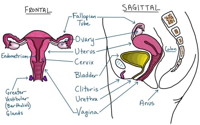
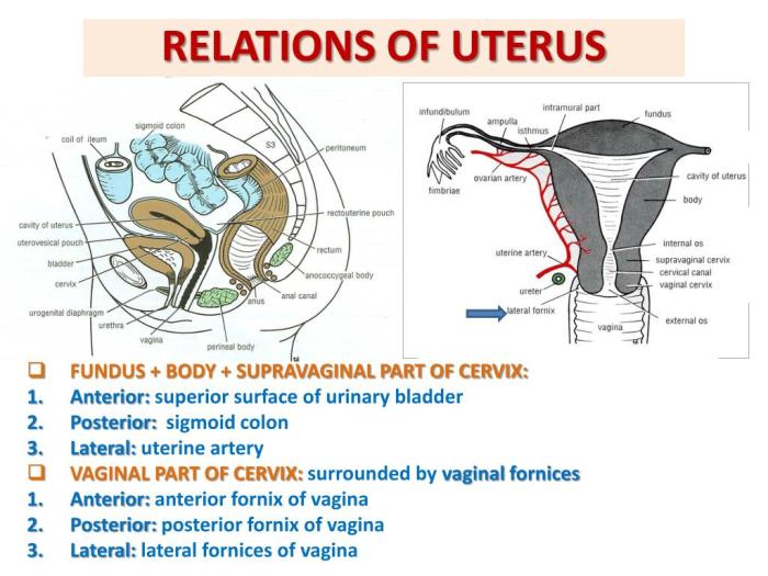
The sagittal view of female reproductive organs provides a comprehensive understanding of the anatomical structures and their relationships within the female pelvis. This view allows for detailed examination of the uterus, cervix, vagina, ovaries, fallopian tubes, and supporting structures, enabling healthcare professionals to diagnose and manage various gynecological conditions.
This article will explore the anatomical overview, key structures, clinical applications, and frequently asked questions related to the sagittal view of female reproductive organs.
Key Questions Answered: Sagittal View Of Female Reproductive Organs
What is the sagittal view of female reproductive organs?
The sagittal view is a cross-sectional image of the female pelvis, obtained by dividing the body along the sagittal plane. It provides a detailed view of the reproductive organs from the front to the back.
What are the key structures visible in the sagittal view?
The key structures visible in the sagittal view include the uterus, cervix, vagina, ovaries, fallopian tubes, and supporting structures such as ligaments and muscles.
What is the clinical significance of the sagittal view?
The sagittal view is used in clinical practice for diagnosing and managing gynecological conditions. It aids in evaluating uterine abnormalities, cervical pathologies, vaginal disorders, ovarian cysts, and fallopian tube issues.


